

For years, endometriosis has been one of the most underdiagnosed and misunderstood conditions affecting women and people assigned female at birth. Characterized by symptoms such as chronic pelvic pain, painful periods, pain during intercourse, bowel or bladder symptoms, and infertility, endometriosis has historically been associated with significant delays in diagnosis. Although awareness has improved, many people still wait an average of 7 to 10 years before receiving a diagnosis.¹˒²
Fortunately, advances in imaging are changing the way endometriosis is diagnosed. Today, transvaginal ultrasound (TVUS) with endometriosis mapping has emerged as a valuable, non-invasive tool that can identify many forms of endometriosis without requiring surgery.³
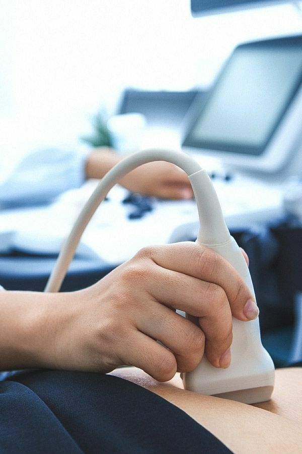
Endometriosis mapping is an advanced transvaginal ultrasound performed by clinicians with specialized training in identifying the characteristic features of endometriosis.
Unlike a routine pelvic ultrasound, which may miss many endometriosis lesions, this specialized examination systematically evaluates the pelvis for signs of:³˒⁴
Most specialists follow the standardized International Deep Endometriosis Analysis (IDEA) protocol, which was developed to improve consistency and diagnostic accuracy across imaging centres.⁴
Research over the past decade has shown that expert-performed TVUS is highly accurate for detecting deep infiltrating endometriosis and ovarian endometriomas.⁵
A large systematic review found that TVUS demonstrates high sensitivity and specificity for many deep endometriosis locations, particularly involving the rectosigmoid bowel, bladder, and rectovaginal septum when performed by experienced operators.⁵
However, TVUS remains less sensitive for superficial peritoneal endometriosis, meaning a normal ultrasound does not completely exclude the condition.³˒⁵ Clinical symptoms and physical examination remain important parts of the diagnostic process.
Historically, laparoscopic surgery was considered the definitive way to diagnose endometriosis. However, international clinical guidelines have evolved.
Current recommendations from the European Society of Human Reproduction and Embryology (ESHRE) support using transvaginal ultrasound or MRI as first-line diagnostic imaging in patients with suspected endometriosis. Imaging findings, together with symptoms and clinical evaluation, can often provide enough information to begin treatment without requiring immediate surgery.³
This represents an important shift in care.
Earlier diagnosis may help reduce years of uncertainty, allow patients to begin symptom management sooner, improve surgical planning when needed, and reduce unnecessary diagnostic procedures.¹˒³
Because endometriosis mapping requires additional training, not every ultrasound clinic performs this examination.
If you’re seeking evaluation for possible endometriosis, consider asking:
Endometriosis mapping with transvaginal ultrasound is changing how clinicians diagnose endometriosis. While it does not detect every form of the disease and does not completely replace surgery in every case, it has become an important first-line diagnostic tool supported by current international guidelines.³
For many people living with chronic pelvic pain or other symptoms of endometriosis, access to expert imaging may mean receiving answers sooner, reducing unnecessary delays, and helping guide more personalized treatment decisions.


Photodynamic therapy is a treatment typically used in the treatment of acne, warts, and a range of cancers, including mouth (oral), head and neck, lung,

The HPV Infection: Do I Have It? But…I’ve never noticed any symptoms so I couldn’t have gotten an HPV infection? Most strains of HPV are low-risk strains that

Are you worried about HPV? Are you unclear as to what exactly HPV is? We at Papillex understand your concerns. HPV can be scary! That’s

Human beings are naturally programmed to eat foods that are healing and beneficial for our bodies. However, we all know that it isn’t always easy
Check out our shop to see what's available
Easy Delivery. Effortless Wellness.
Subscribe for:
By signing up for a Wellex subscription, you authorize use of the credit card you have on file and shipping at the frequency you have selected. Rest assured you can change or cancel at any time.